How do you go about capturing images of a coronavirus, which is too small to see with a standard light microscope? Here’s a 5-minute video in which Vox explains how two electron microscopy techniques give us views of what the SARS-CoV-2 virus looks like.
The SARS-CoV-2 virus measures around 100 nanometers, meaning 10,000 virus particles can fit into one millimeter. Since the shortest wavelength of visible light is around 400nm, we can’t capture photos of the virus with visible light.
To capture images of smaller things, scientists accelerate electronics in a field to cause them to behave as waves with a wavelength much smaller than visible light.
“Two electron microscopy techniques, SEM and TEM, offer different views,” Vox writes. “A Scanning Electron Microscope (SEM) scans the surface of a sample and records information that bounces back, similar to a satellite image. A Transmission Electron Microscope (TEM) transmits electrons through a sample and projects a cross section of its inner structure.
“Together, these images help scientists observe the virus and how it moves in and out of host cells.”
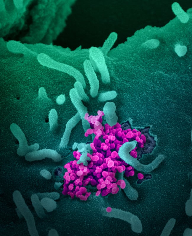
Images captured with both techniques come out in black-and-white, and color is added afterward for clarity. Here are some example images captured with the two methods:
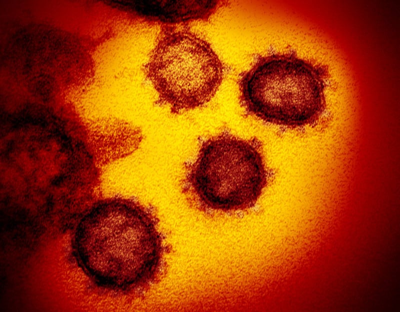
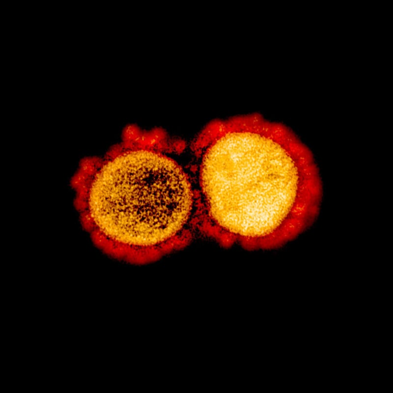
Transmission Electron Microscope (TEM) Images of SARS-CoV-2


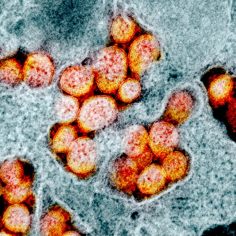

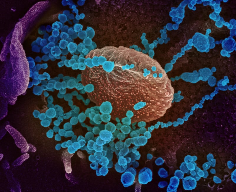
Scanning Electron Microscope (SEM) Images of SARS-CoV-2


You can find more electron microscope images of the coronavirus in the NIAID album “Novel Coronavirus 2019” over on Flickr.
Image credits: All images by NIAID


